
Nova Derma | Amman, Jordan
Mole Check & Skin Cancer Screening in Amman
Dermatologist Assessment of Moles and Changing Skin Spots
Check the mole before deciding how to remove it.
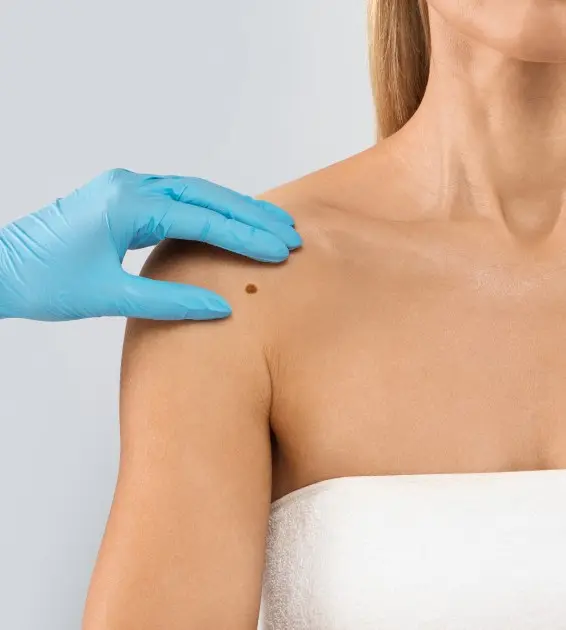
Most moles are benign, but a new or changing mole can sometimes need closer assessment. At Nova Derma, moles and suspicious skin spots are evaluated clinically and with dermoscopy when appropriate.
Not every mole needs removal or biopsy. Some moles can be monitored safely, while a mole or skin spot that remains suspicious may require biopsy or excision for histopathologic examination.
Warning signs
The ABCDE Warning Signs
ABCDE is a guide to what to look at, and it is worth checking your moles and skin spots against it.
- A — Asymmetry: one half of the mole does not resemble the other half.
- B — Border: the edge is irregular, notched, scalloped or poorly defined.
- C — Color: there are different colors or uneven pigmentation within the same spot.
- D — Diameter: melanomas are often larger than about 6 mm when diagnosed, but they can be smaller.
- E — Evolving: the mole is changing in size, shape, color or symptoms over time.
A change does not automatically mean cancer, but it is a reason to have the spot assessed rather than watched indefinitely.
ABCDE is a useful warning guide, not a self-diagnosis tool. A suspicious spot should be assessed clinically.
Who May Benefit From a Mole Check?
The need and interval for future skin examinations are individualized rather than the same for everyone, so the assessment is built around your own moles, your history and how your skin has changed.
A mole check is worthwhile if you
- Notice a new or changing mole
- Have many moles or atypical-looking moles
- Have a personal history of skin cancer
- Have a strong family history of melanoma or skin cancer
- Have significant previous sun exposure or repeated sunburns
- Are worried about a skin spot that looks different from the rest
Removal may need to be modified, deferred or referred if there is
- A mole that is clinically uncertain, because a suspicious or uncertain lesion should not be destroyed with laser, electrocautery or another tissue-destructive technique before histopathology has been decided
- A mole that is not suitable for in-clinic removal, which may need surgical assessment instead
- An inflamed, infected or traumatized area, which should settle before the skin is assessed
- A lesion in a location where biopsy would be complex, which is referred rather than treated in clinic
How a mole is assessed
Clinical examination: the dermatologist looks at the mole's shape, color, borders, symmetry, location and whether it is changing. Your personal and family history may also influence the level of concern.
Dermoscopy: dermoscopy uses magnification and polarized light to examine structures and pigment patterns that are not easily seen with the naked eye. At Nova Derma, dermoscopy is available as part of the assessment of selected moles and pigmented lesions.
Follow-up when appropriate: if a mole appears clinically suitable for observation rather than immediate biopsy, the dermatologist may recommend follow-up examination and comparison over time. The timing depends on the individual lesion and risk profile.
Biopsy or excision when needed: if a mole or skin spot remains suspicious, biopsy or complete excision may be recommended. The removed tissue is sent to an external pathology laboratory for histopathologic examination.
Not every mole needs a biopsy. Biopsy is used when the clinical assessment indicates that tissue diagnosis is needed.
Can benign moles be removed?
Yes, selected benign moles can be removed for symptoms, repeated irritation or cosmetic reasons after appropriate assessment.
The removal method depends on the mole type, depth, location and whether histopathology is needed. A mole that is clinically uncertain should not be treated as a purely cosmetic lesion.
What happens if a biopsy is recommended?
- The area is numbed with local anesthesia.
- The dermatologist removes the appropriate amount of tissue based on the clinical question.
- The specimen is sent to an external pathology laboratory for microscopic examination.
- Further management depends on the histopathology result and whether any additional treatment is needed.
Nova Derma does not operate an in-house pathology laboratory.
Most moles need nothing more than observation; the aim of the check is to identify the minority that need tissue diagnosis.
Red flags
Which Moles Should Be Checked?
A mole deserves assessment if it is new, changing, looks different from your other moles, or develops new symptoms.
- A new mole or dark spot that looks unusual
- A mole changing in size, shape or color
- Irregular or poorly defined borders
- Multiple colors within the same mole
- A mole that looks different from the rest — the “ugly duckling” sign
- New bleeding, crusting, persistent itching or tenderness
- A spot that keeps changing or does not heal as expected
A change does not automatically mean cancer, but it is a reason to have the spot assessed.
A suspicious lesion is not destroyed first
Laboratory testing is arranged externally
Your dermatologist
Moles Assessed by a Specialist Dermatologist
Dr. Qusai Shakhatreh
Specialist in Dermatology & Venereology
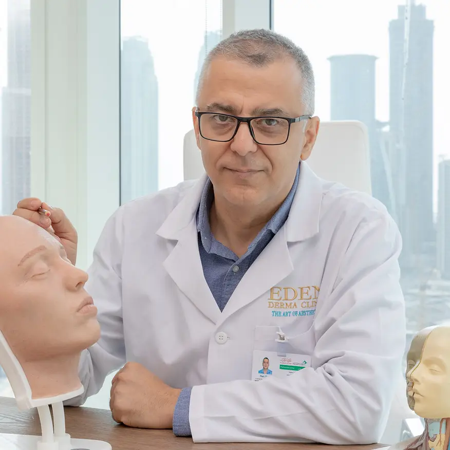
A mole is assessed clinically first, and dermoscopy is used where it adds to the picture. Dr. Qusai Shakhatreh, Specialist in Dermatology & Venereology, examines the mole and your history, and decides whether the lesion can be monitored safely, needs a follow-up comparison, or should go to biopsy or excision so the tissue can be examined histopathologically.
Mole Check FAQ
Does every mole need to be removed?
No. Most moles do not need removal. Some can be monitored safely, while suspicious or symptomatic moles may need biopsy or excision.
Does every mole need a biopsy?
No. Biopsy is recommended when the clinical and dermoscopic assessment indicates that tissue diagnosis is needed, and not as a routine step for every mole.
What is dermoscopy?
Dermoscopy is a non-invasive examination that magnifies and illuminates a mole so the dermatologist can assess pigment and structural patterns more closely.
Can dermoscopy prove that a mole is benign?
Dermoscopy can improve clinical assessment, but when a lesion remains suspicious, histopathologic examination after biopsy or excision may still be necessary.
Can I have a mole removed with laser?
A mole should first be diagnosed. If it is suspicious or if histopathology may be needed, laser, electrocautery or other destructive treatment should not be used instead of obtaining tissue for examination.
What if my mole has changed?
A changing mole should be assessed, particularly if there is evolution in size, shape, color, border or symptoms.
Are large moles always cancerous?
No. Size alone does not diagnose melanoma, and melanoma can sometimes be smaller than 6 mm. The overall clinical pattern and change over time matter.
Can you remove a benign mole for cosmetic reasons?
Yes, in selected cases after assessment. The technique depends on the type, depth and location of the mole and whether tissue examination is appropriate.
Do you have a pathology laboratory at Nova Derma?
No. If a biopsy or excision is performed, the specimen is sent to an external pathology laboratory for histopathologic examination.
A mole check is a clinical review, not a treatment appointment, and it often ends with monitoring rather than removal.
Concerned About a Mole or Changing Skin Spot?
If a mole is new, changing, looks different from your other moles or simply concerns you, a dermatologist assessment can determine whether it can be monitored safely or whether biopsy or excision is appropriate.
Nova Derma · Amman, Jordan